When checking the patient’s identity after arriving at the room, a wireless barcode scanner* allows you to freely approach the patient and scan information without being restricted by obstacles that may be near you and the patient. With a wireless hand switch that supports up to 10 m (33 ft) range, users can take images from far away to reduce radiation exposure.
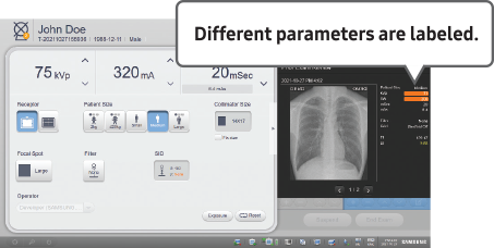
Since the role of mobile DR is usually to use follow-up exams, it is important to perform x-ray exams under the same conditions. Prior Exam Review* displays previous images and exposure parameters of the patient being examined. With a quick comparison, users can improve image consistency and reduce retakes.
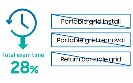
SimGrid™ application streamlines the workflow by guaranteeing image quality without the use of a conventional grid. This allows the omission of grid installation and removal steps from the conventional workflow leading to 28% reduction in total exam time.
In portable exams, radiologic technologists rotate images after the acquisition as the image orientation depends on the exam environment. Image Auto rotation* detects the rotation angle of the chest/abdomen/pelvis/infantogram image and automatically rotates in the correct direction based on AI algorithm. The auto-run option of advanced applications creates the companion image without additional settings or x-ray exposure.
In the ER/OR or Trauma environments, multiple staff may often need to quickly acquire and check images in real-time. Mirror View* provides secured screen sharing for healthcare team to review images. This allows staff to check images together on a separate screen, reducing the time to first aid and the risk of contamination. Through QuickLink, you can check whether the acquired images uploaded well to the server by accessing RIS/HIS directly without a separate PC.
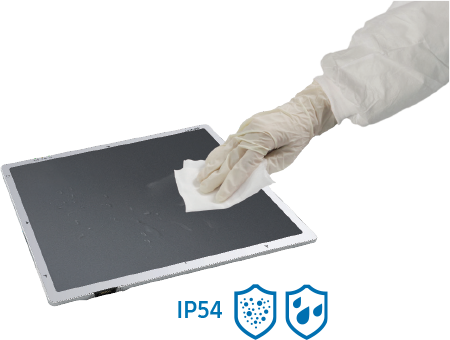
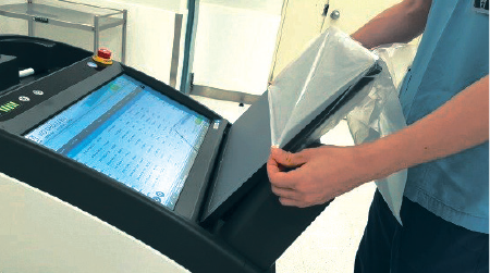
When preparing for the next exam, EasyClean, which locks the screen for a while, helps you clean equipment quickly and control infection effectively.

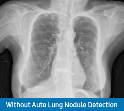
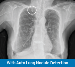
Auto Lung Nodule Detection is computer aided detection software to identify and mark regions in relation to suspected pulmonary nodules from 10 to 30 mm in size. It is designed to aid the physician to review the PA chest radiographs of adults as a second reader and be used as part of S-Station.
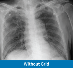
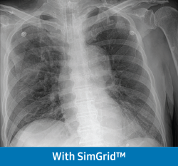
With just a click, SimGrid™ allows you to provide better patient care with higher satisfaction and reduced retake rates without the use of a portable grid. It improves image contrast by reducing scatter radiation effects and creates better image quality. The 3-step intensity control (Low/Medium/High) enables customized image processing.
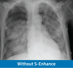
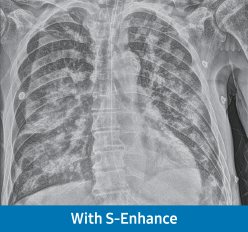
To support your diagnosis, S-Enhance improves the clarity of foreign bodies (e.g. tube, line and/or needle) in images of chest, abdomen, and L-spine. With a single on-screen click, the companion image is created without additional settings or x-ray exposure, streamlining the workflow.
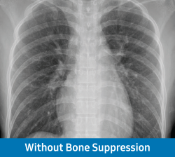
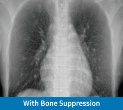
Without additional setting or exposure, Bone Suppression Imaging improves the clarity of soft tissues by suppressing the appearance of bones in chest
images, which improves your ability to detect nodules. You can easily create the companion image with just a click on the screen.

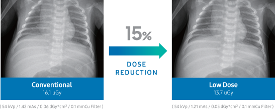
Underaged patients are more radiosensitive than adults. To alleviate these concerns, the new S-Vue™ engine helps achieve the optimal dose level for children during pediatric x-ray scans. The dose level can be reduced up to 45% for pediatric abdomen, 15.5% for pediatric chest, and 27% for pediatric skull exams. This is especially significant as abdomen protocols may include genital regions2.

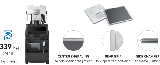
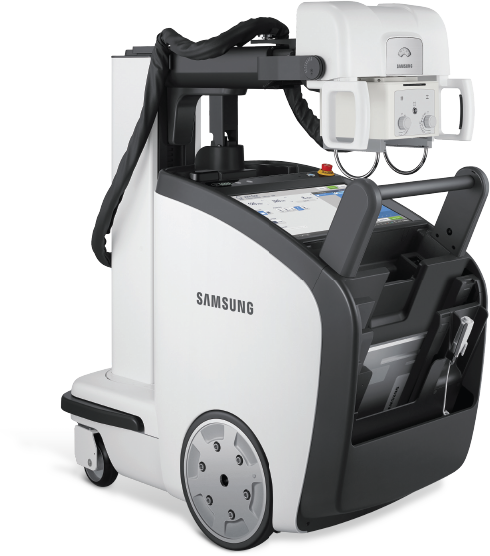
Ultra-light GM85 Fit provides an effortless driving experience for users. Not only the equipment but also the detector that must be held directly with the arm should be light. The user-centric design of AccE Standard Detector supports patient positioning and alleviates daily burdens.